 Discovering something means getting knowledge of something one did not know before. For the discoverer himself this may lead to rather strange experiences. Most discoverers are usually like any human being, however, their curiosity and ability to imagine the appearance of the piece that will enhance our knowledge of life are very distinguishing features. Often they have also kept the ability of the child to absorb new knowledge.
By Dr Erik Enby (artikeln på svenska) | Homepage: Enby.se | Mail: erik [at sign] enby.se | Translation by Birgit Oreskog and Siv Wernborg
Many great researchers have said that one must be like a child to be successful in the world of discoverers and scientists. This does not mean that such a person is childish and naive. Instead it is a question of how to take an innocent attitude to those around, to be open and unprejudiced. Such individuals, therefore, may seem somewhat naive when in their eagerness they try to tell what they are doing and inform about their findings. Sometimes they can suddenly seem almost mentally disturbed.
How did Albert Einstein (1879-1955) look when he discovered that a door can be made to open by breaking a ray of light? Or imagine the reaction of Wilhelm Röntgen (1845-1923) when he realized that it is possible to see right through an organism! Imagine Alexander Fleming (1881-1955) when he realized that infections can be cured with something that was later named penicillin!
It often happens that the discoverer cries: ”Look!” Others look but understand perhaps nothing, and the discoverer remains alone with his discovery. So far he himself does not know much, because he sees what he has discovered for the first time. The discoverer usually has not led the life of an ordinary person, because then he would not have made any discovery. As everybody knows, there is something called ”being out exploring”.
One has to do that in order to find something new. When the explorer makes his discoveries he is astonished, but perhaps he says: ”Wasn´t that exactly what I believed?” Already he had an idea of what he was after, because otherwise he would not have embarked on his expedition. It is usually in connection with such a mysterious inner force that the explorer makes his discovery. The question is if this can be called curiosity, and if there is at all any word for the quality of being this way. He feels what is out there in the unknown, and suddenly he finds it.
How this happens is a mystery, but it takes place in his mind. It is really like doing a jigsaw. All the pieces are there. Some are in place, but it is a question of finding the remaining and put them together with the rest in order to reach the optimal result. This can take a very long time.
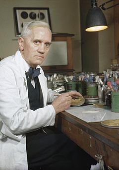 What we call chance sometimes has had a great importance in such a connection, because it was certainly chance that made Alexander Fleming discover the penicillin. That this happened just to Fleming, of course, was a consequence of what he was working with as well as the conditions prevailing in and round his laboratory. When he returned to this from a vacation he noticed that in some areas of an unwashed culture of bacteria there were no bacteria growing.
Alexander Fleming – Photo: Wikimedia Commons – Imperial War Museums
Fleming noticed this being no result of an experiment of his, but something that showed itself to him by coincidence – chance – and could as well have avoided his attention. Fleming, however, was observant enough to notice the phenomenon, and as he understood that there must be a reason for this, he started looking for it, and you can say that this work was a consequence of luck.
The fact that Fleming in that way was able to explain that a penicillin producing mould was the cause of the phenomenon, on the other hand, was no coincidence – chance. If the presence of this fungus could be explained by the fact that a colleague next door was working with fungi, the spores of which may have found a suitable soil for their continued development to the penicillin-producing bacteria-counteracting fungus in Fleming´s unwashed culture of bacteria, is something one can just suppose, but this must then have been coincidental. The circumstances thus opened the doors for Fleming, who thanks to being unusually lucky as well as observant realized what he should do in his laboratory. This resulted in Fleming receiving a Nobel Prize.
Sometimes the discovery happens in a dream. This is told by professor August Kekulé (1829-1896), who claimed that in a dream he had experienced something telling him that the 6-membered ring in benzene existed. How he later on showed that this was the case, is another story, however, it is told that, thanks to what happened in his dream, he ended up right in his work. Frédéric Chopin (1810-1849) had paper and pencil beside him when sleeping, because suddenly he was inspired in his sleep. At once he wrote down what he had dreamt, and it is told that, among other things, the revolution étude was made this way.
Finding the place where the Inca treasure lies hidden could perhaps happen in a similar way. Perhaps the place would show itself before one´s eyes if, during a very long period of time, one would occupy oneself intensely with finding the treasure. However, only those who try hard can experience something like that, and discoveries mostly are made only by persons who exert themselves to the utmost.
Another way of inventing can be rediscovering something that has been known before, but has been forgotten, or for different reasons has escaped arousing interest, or has even been kept secret. An example of this is the rediscovery of the continental shift, which was imagined already 300 years ago, and which in the beginning of the twentieth century was mentioned again by Alfred Wegener (1880-1930). Gregor Mendel´s (1822-1884) research in the theory of heredity (Mendel´s laws) is another example.
Myself I have experienced some of all this. I have already mentioned the expedition. But why did I embark on it? Well, there is a reason for everything, and the reason was that there were things happening around me, which I wanted explained.
Early experiences of disease awakened my interest for medicine
I was young when I noticed that illnesses were an evil in people´s lives. My brother fell ill when he was about fifteen with a serious illness, which, after a long period of suffering, caused his death. Any acceptable explanation of his illness could not be provided by the doctors. I understood that, if you don´t know what causes an illness, you cannot normalize the sick person. The other thing that determined my decision to explore was that Gunnel – my future wife – in the course of four days was almost completely and definitely paralysed. She was sixteen years then, and the sickness led to a lifelong suffering in a wheel chair.
Suddenly I was surrounded by sickness, and it became impossible for me to behave as though nothing had happened and not to bother. What had happened was to become of thorough and decisive importance, both to those directly affected and to the next of kin. It did not result in me directly hating sickness, but it worried me that nobody knew anything about this phenomenon. There was simply nobody who could answer the question how a person´s long life, owing to disease, could change so completely. Why didn’t anybody have a constructive knowledge of the phenomenon sickness? What happened in the tissues? Why were some of them defenceless, others not?
Even if the problem of disease was new to my family, people have had disease problems since millions of years. It kept worrying me, and I dare say that was the reason for my early decision to try and get to know the essence of diseases.
Of course I would choose the profession that means occupying oneself with illnesses of all kinds. This was obvious considering my level of ambition. So after 12 years I was a doctor with two specialist competences and the right to work independently with the illnesses. However, I had not at all found out what made a chronic disease slowly showing signs of its presence, being able to disturb the sick person for years, and that was exactly what I wanted to know.
When I saw that I could not find the answer within the walls of the hospital, nor could I find a colleague to talk with about this matter, I started my expedition. This meant that I shook off all of the cramming studies necessary to continue the medical career in order to go on developing myself freely instead. I declined all offers of a doctor´s thesis concerning something that did not interest me and began more and more looking upon the hospital as a supporting institution. There it was not possible to learn to know the ill health conditions the way I had in mind. I was not allowed to do research the way I wanted and there was no supervisor who would help me on my conditions.
The first part of the journey meant only browsing in the scientific world looking for anything that could be interesting. What was it that kept an individual sick and slowly destroyed his body? This took rather a long time, and not until I studied the diseases of the vegetable kingdom, I started to have a feeling of how the chronic complaints of man begin. The collected works on the reasons for the diseases written by the French Professor Jules Tissot, published in 1926, definitely put me on the track: ”Constitution des organismes animaux et végétaux. Causes des maladies qui les atteignent”. (Constitution of animals and plants organisms.Causes of diseases attacking them.)
I had noticed that the plant diseases had always had a connection with a fungus attack, destroying different tissue parts of the plant and that this could happen at a varying speed. Tissot thought that it can be in the same way with tissues from animals and human beings and had even written a whole chapter about the development of the fatal disease cancer having a connection with a fungus attack. It was then that I began to suspect that the tissues are broken down in a fundamentally similar way in all nature, it may be a question of plants, small and big animals or humans. A chronic, protracted sickness could have something to do with different forms of attacks. So, I allowed myself to extrapolate this theory.
In nature the overripeness comes after the ripeness, and then always the putrefaction, if everything is allowed to have its normal course. This process of destruction of the ripe seems to be something completely normal and is always part of the life process. When I looked upon this in the microscope I noticed different kinds of vegetation in tissues, which were on the way to decay. In my investigations of fruit and vegetables in the breaking down phase it was never a question of an inexplicable decay of tissues. Different kinds of growth were going on in these and caused the decay.
This vegetation could easily be studied. One just had to put a lemon or an apple showing the first signs of destruction, under a glass cover and study the continued course of destruction. Under the cover one could then see that there would always be a mould carpet growing on the whole fruit, and if things were allowed to take their course, then the fruit was nearly completely destroyed in this connection. Especially interesting was performing the experiment with a citrus fruit which, after about two months, had disappeared almost completely under the cover.
Thus, studying plant diseases was rewarding. Seeing how normal ripeness was transformed into normal putrefaction was as interesting as studying a disease in a plant. In both cases there was a change and destruction of the tissue of the plant as well as of the fruit by something looking like a fungus process.
With insects the case is similar. Even such creatures can be ill, and if they are examined one can see that they are almost always exposed to some kind of fungus attack. A better known attack is the so-called fly mould which kills flies in the autumn. Other insects have their special attackers, and fact is that hundreds of thousands of different forms of fungi are known, which can grow in and destroy tissues of vegetable as well as animal origin.
A fact which I have found very interesting when I began studying the destruction of tissue, especially in the vegetable field, was that the growing of fungus is not taking place anywhere in nature. This may be hard to understand for a completely uninitiated person, however, considering that the chanterelles belong to the juniper hill and the mushrooms to the field where the cows go, it will soon be easier to understand what I mean. If you look for mushrooms in the juniper hill you will not find any there. Most people know that. With the mould fungus plant it is about the same. The mildew growing on the gooseberry bush keeps there but does not grow out on the currant bush nearby. Neither does the mildew destroy the gooseberry itself but is only growing on its surface, leaving the berry otherwise in peace. It is quite possible to eat the berry after having scraped off the mildew. It is here a question of a superficial so-called mycosis which keeps itself only on the surface of the berry.
Nor does the hated potato rust fungus spread to the carrots or the radishes, but keeps itself to the potato plant. Unlike the mildew it grows into and destroys not only the plant above the soil, but sometimes even the potato. Growing a new sort of potato next year can turn out well, but if one chooses to grow the same sort again, there may be rust attacks again. Fly-mould does not attack grasshoppers or wasps. There is thus no risk of infection for these insects even if all flies get sick and die because of fly-mould infection.
Thus, in nature there is no chaos, but orderliness. Everything grows only where it can grow, and if there are not the right growing conditions, nothing will grow. It does not matter how much spores are blowing to and fro and are taken by the wind to all thinkable places. Only if they land in an environment favourable for them, they can root and begin to grow.
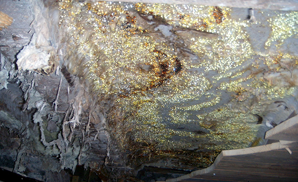
Serpula lacrymans – Wikimedia Commons
A very remarkable phenomenon, which made a very deep impression on me during my expedition, was that it looked as if all these moulds with their spores in some way lay in wait everywhere in the nature, to begin to grow as soon as they had a chance. An example of this is the so-called dry rot fungus which starts its destroying activity as soon as humidity has forced its way in a wooden construction. It is also opportunistic in so far as if it can only send out a few runners down the foundation, so that it can come down to the water needed for its growth, then in a period of ten years it can destroy a whole wooden house. It is no overstatement to say that the underlying process that made the wooden construction of the house rot, was very slow. A way of preventing further destruction is removing all moisture. Then the fungus cannot grow further and destroy the wood it was about to grow into.
That could be done with the dry rot fungus, but not if a similar fungus-growth destroyed a tree in the forest. Even if big parts of a tree trunk were destroyed by a mould fungus inside, the tree could yet look fresh and fine, growing in the forest. How could this be, and why did it happen? Well, a complete answer to that question I could not find, but I had understood that different growing conditions must prevail to make it possible for these fungus attacks to start.
Now it was time to start thinking about the condition of man – the living creature who suffers from so much sickness. Could it be with man as with the rest of nature when illnesses are concerned? Were these a consequence of plant attacks of different kinds? I went on extrapolating, and based on everything I had now found out about the sicknesses in the world of animals and plants, my theory was that it could be that way.
But science claims that what is presumed in a hypothesis must be proven to be right, otherwise, however interesting it may be, it is and remains just a theory. I decided to investigate the chronically sick microscopically in order to find out in this way if there existed some form of vegetation in their blood and tissues.
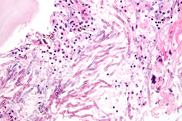
Micrograph of showing a fungal pneumonia (pulmonary aspergillosis). H&E stain. Wikimedia Commons
Somebody now says that such a work has probably already been done by someone else. The scientific field is rather large and microscopical work was not new. It is quite right to expect this and in order not to risk to a great extent doing what others have done, you have to make a thorough research. What was the earlier result from the field to be investigated?
I found that my ideas concerning the states of disease belonged to a tradition with roots in early French and German microbiology. Advocates as for instance Claude Bernard (1813-1878), Antoine Béchamp (1816-1908), Jules Tissot (1870-1950) and Günther Enderlein (1872-1968) a long time ago had the idea that chronic illness could possibly be caused by attacking microbiological processes in the tissues. When I studied their works concerning the underlying causes of the development of chronic illness, I noticed that my ideas in the matter agreed well with theirs. It concerned different forms of vegetation in the tissues, irritating and slowly changing them.
During my medicine studies, or in my work in the hospital, I had never heard of anything like that. Either this was completely wrong, or, for reasons that I could not understand, this trace had been neglected by scientists who preferred looking for the causes of illness among the chromosomes of the tissue cells and disturbed biochemistry. The last-mentioned, however, could never catch my interest, as I preferred to see illness as the result of an attack on the organism. Now I started to think that there were enough reasons for investigating if there could be any truth in this. So I started the microscopical work.
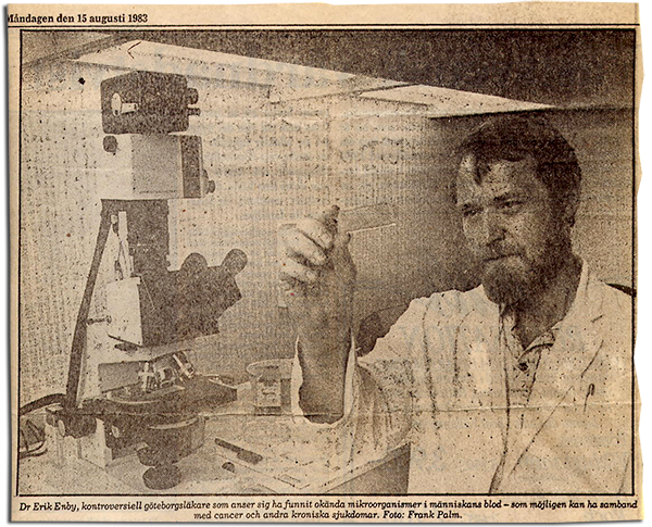
From the article: ”Okända maskar i blodet” (Unknown ”worms” in the blood) i GP August 15, 1983 – Photo: Frank Palm
To begin with this meant just investigating the blood from chronic invalids. Was there anything in this that did not exist in the blood from healthy individuals? Healthy blood was described in the hematology literature, which says that blood is always sterile, which must mean that, beside red and white blood corpuscles and thrombocytes, there is normally no growth in it at all. Therefore I was very surprised when in the blood from my chronically sick patients I often could find a massive presence of particles that did not at all appear to the same extent in the blood from healthy individuals. In the beginning these particles seemed to be a mess of small non-uniform formations spread among the red corpuscles. The question now was how to react to this phenomenon.
Were these particles a normal discovery or not? Did they consist of just small fat balls, or could it be that here among the corpuscles there existed a presence of micro-organisms? The only possibility of getting some clarity on this was finding out if the particles were living or not. Fat balls show no signs of a life process, but micro-organisms on the other hand would be able to do this. Culturing would be one way of settling this, but I had no possibility of isolating these particles to see if they were growing in a culture. Nor would it be possible to somehow get these particles to multiply on a culture medium outside the body.
I decided for the time being just to look at all this to see if anything would turn up in the blood tests that would explain the existence of the particles. Today I have investigated blood from thousands of patients and have just to look very briefly to judge if it is looking alarming or not. It surprises me that 30 years ago I went on looking at all these particles in order to get an explanation why they were there among the red corpuscles. Anyone would have given up and realized that it would be impossible to get all this “jumble” appearing in the microscope in order.
What called my special attention was that the particles did not look quite the same at the different states of illnesses. Looking at them in the maximum magnification, one could see that they often crawled around among the corpuscles, that they had their own spontaneous movement. It was possible to follow them in the microscope for almost a week, if the blood did not dry out. They not only moved in the slides but could sometimes also change their form into small worm-like structures. This could not happen if they consisted of small fat balls.
By looking at capillary blood from all the chronically ill people I could get into contact with in the surgery, I got so much information about all these particles that I was more and more convinced that they represented a living material. I looked and looked, and when patients with unusual sicknesses turned up I looked for new pieces in the blood tests. From where did all these particles come and what became of them? I was pretty sure that there had to be a production of these particles somewhere in the organism, in the same way as when a flower creates pollen.
An experience I have made in my life is that if you don´t give in, then the help will come suddenly from an unexpected source. It was necessary that something like that would now happen. I began to doubt if it was possible to investigate all this, in any case the way I was trying. Was it possible to go any further?
Particles and small flake-like structures growing in volume
Then suddenly a mother with her newborn and retarded child made a visit. The child completely lacked steadiness, seemed half dazed and hung like a clincher on her arm. Of course the mother understood that there was not much I could do for the baby, but perhaps its general state of health could be improved? As usual I made a vital blood microscoping and found a massive quantity of particles vibrating in a speed that I had never seen in a blood sample before. This was something completely new. Moreover, among the corpuscles there were many small flake-like structures. I had seen those before in the blood tests, but not yet given them any attention. They could look differently but lay there motionless in the blood, looking rather uninteresting.
Now I could suddenly see how such a flake could start a production of small particles looking like those that could be seen among the corpuscles. This thus took place outside the child´s body and could be seen during the microscopical analysis of the blood. The particle production of the flake was very intensive, and myriads of particles, pushing aside the corpuscles around, were produced by this flake during the ten days I could observe it. So, between the microscope slide and the cover glass in the microscope a big pile of granules round the flake was formed.
This was of course a discovery, and if anything a proof of the fact that both the flake and the particles were made of living material, of which probably nobody had written a single word in the medical literature. Thus a very important piece in my work. Now I could extend my theory further and make new assumptions.
The big heap of granules round the whole flake consequently grew and in the meantime it was possible to follow what happened in the microscope. I wondered how long this production of granules would be able to continue, and if such a production was going on in the body of the chronically ill person as well. Therefore I started looking carefully at these flakes when they turned up in the blood tests and found that signs of such a production could sometimes be observed directly in connection with the taking of blood tests.
Another strange fact was that the granules round these flakes increased their volume each day and sometimes changed into small warm-like, movable forms. More could not be seen in the microscope, but this was enough. Now I had at last got an explanation/evidence why there was a massive finding of differently formed particles in the blood from chronically ill people and that some probably were unfamiliar to a healthy blood.
Of course I was tempted to believe that what I had discovered could have something to do with the underlying process of chronic disease. At the same time I was well aware of the fact that the discovery so far meant just that I had come across something unknown. I had not proved that the particles and the flakes were the cause for developing a chronic disease. It was, however, possible to analyse what I had found and in this way understand that the flakes and the production of particles could generate sickness.
Arguing this way, I took it for granted that the flakes were living and could produce myriads of particles if they existed in such a phase and environment that the production could start. If it could start in the circulating blood this would mean that the blood plasma could be totally soiled with these particles, something I have been looking at for years. A feeling of illness could arise successively, perhaps because all these particles went on developing slowly in the blood plasma, changing its chemistry, because growing changes the soil, in this case being the blood plasma, but perhaps also the corpuscles and other tissues.
A flake could start its production of particles at the place, in the solid tissues, causing for instance bleedings, if the production should take place in a vessel wall. Symptoms from the central nervous system could result if a corresponding production of particles could arise in this. If the production of particles did not end, the result could be a change of consistency or an increase of the volume of the tissue, and later this could have to make place for even larger growing material. If it was growing in the same way in the tissues of the sick person as in the microscope, which could be seen in this, it could thus explain rather much of what is happening to the body when this is not quite in control any more – i.e. when the individual feels a kind of vague and protracted strange physical experience – sickliness. However, I could not prove that this was the case, but the expedition was to go on anyway.
The thought that the particles in turn could grow and result in yet another interesting phenomenon in the blood, was something that was natural to think about. And from where did these flakes come? How did they start to grow in the organism? This was certainly a mystery, and it was very clear to me that probably it would not be possible to culture them. Finding out their origin had to be done in a different way. For the time being I had to look upon them as small “plants” in the organism, with their seeding the way that I had been able to see in the microscope. I therefore went on studying the particles, which would take me further a part of the way.
Now I will try again to make a long story short. My blood examination was carried out by letting a drop of blood spread out into a thin film between a cover glass and a microscope slide. As blood has a very low viscosity, everything there was in the blood drop happened to lie side by side in just one layer. The contents of the drop of blood that would otherwise not be possible to see, were then possible to study in the microscope. In the blood from the chronically ill, in the thin layer of corpuscles and blood plasma, there were here and there areas that showed a seed of almost completely destroyed – attacked – corpuscles mixed with the strange particles in different forms and sizes. Also they seemed to keep together and form something that you would call a focus of infection in the drop of blood. A number of such focuses could sometimes be found if the drop of blood was microscoped in its entireness. The corpuscles lying around could look completely normal. The destruction of corpuscles thus kept to limited areas in the capillary blood spreading, and between these areas the corpuscles looked normal in the microscope.
Of course I asked myself if the particles caused these swarm-like focuses in the blood drop. I imagined that the different forms of particles and the destroyed corpuscles were kept together in a small clot of phlegm in the plasma, which could explain that, between these focuses in the blood, there was no destruction of the corpuscles to be seen. The destruction of these thus kept to limited areas, and in other respects the corpuscles looked normal in the microscope. At chronic diseases the capillary blood thus does not consist of a homogeneous mixture of its elements in the plasma.
I now imagined that the consequences of such a formation of focuses also could arise in the solid tissues of the organism, which could then be changed in a similar way. In this case the whole world of particles that I had studied would be able to cause the development of lots of symptoms which mysteriously occurred at many unclear chronic states of illness.
When the blood flowed out to a thin film between the cover glass and the microscope slide, everything that seemed to be strange for a healthy blood could therefore easily be studied in the microscope. In order to get a reasonably right idea of all this, one has to imagine how this strange thing seen in the blood film looked in the blood drop before this had flowed out to the thin blood film between the glasses. Thus, one had to think backwards to understand how the unknown structures in an almost two-dimensional level of capillary blood from chronic invalids looked when they existed in the three dimensions of the untouched capillary blood. This was the only way to understand how this vegetation could be able to form lumps in blood and in solid tissues, where they could probably reach considerable sizes before it was noticed what could be the consequence – the symptoms – of the different forms of growth apparently going on in blood and tissues at protracted chronicity.
Examining blood under the microscope now and then revealed, in large cutouts in the blood spread, areas of what appeared to be lifeless material. If they were enlarged, one could see that this material looked like a completely dead moon landscape. Extremely strange was the fact that, in the periphery of such cutouts, a rather broad zone of moving particles, varying in size and form, could always be observed. At the different chronic diseases that I examined, one could, when carefully observing these formations, see that in principle they were built in the same way, but that they differed in detail. The building stones in the seemingly lifeless material and the forms and sizes of the movable particles varied.
This discovery in the blood film must, in the untouched drop of blood, exist as a ball-shaped formation with a peripheral zone/area, consisting of tens of thousands movable particles in different sizes and forms. Owing to the form that this material showed in the microscope, I called these discoveries “disc-formed regions with a peripheral fringe area”, even if these regions probably existed as ball or bullet formations in untouched capillary blood. Their existence in different sizes indicated that they were growing, and claiming that these balls were made in the body of the sick person was reasonable. I could not imagine that these ball formations could grow in a culture. No, to me it was obvious that the growing up of such “balls” could only take place in an organism increasingly falling ill. The growth zone was the peripheral zone/area of particles with front against the surrounding, not yet destroyed tissue.
Repeated examinations under the microscope of patients with such a growth of balls indicated that this happened very slowly, which could explain the tendency of the chronic illness to be protracted. I assumed that this growing principle would take place in the solid tissues of the organism, which could then be damaged and cause symptom pictures. All these growing principles: flakes, swarms and ball formations could exist at the same time in the capillary blood, especially if the illness of the chronically ill person was far advanced.
The way of taking a test with a pinprick in the pad to get capillary blood results in this being mixed with tissue fluid and tissue material from the tissue lying between the capillaries. The focuses to be seen together with the conglomerate of corpuscles in capillary blood from the chronically ill person thus do not reveal anything sure about where in the tissues/somatic architecture these focuses are growing up/developing.
The lumping of blood cells is an important discovery
Another matter that was strange in the capillary blood of the chronically ill was that the corpuscles tended to cluster together to large heaps. Sometimes this could be so far advanced that, at the examination of the blood under the microscope, there was not a single free blood corpuscle to be found.
The blood thus formed lumps, and this phenomenon could be compared with what is happening when different milk pathogens – lactic acid bacteria – affect fresh milk. This will then become sour in different ways, but there will also be a forming of lumps or conglomeration of the contents of the milk liquid and there will be yoghurt, soured whole milk, ropy milk, or what used to be called sour milk.
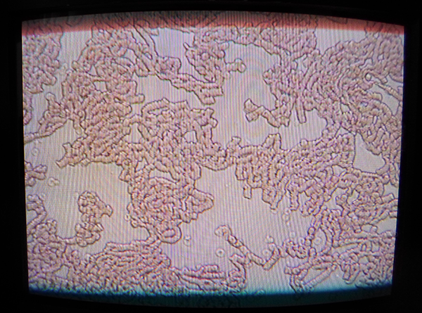
Dr Erik Enby has found lumping in a patient’s blood – Photo: Torbjorn Sassersson, NewsVoice.se
The lumping phenomenon arises in blood from anyone with chronic diseases. Its viscosity then increases and the circulating speed will quite likely, from a general point of view, be lower in the whole organism.
Professor Melvin H Knisely (1904-1975) was the very first one to investigate this condition which proved to occur more or less at all chronic diseases. Professor Lars Erik Gelin at Sahlgrenska University Hospital in Gothenburg confirmed in the fifties the result of research of Knisely, and in the beginning of the sixties this was done also by V. O. Björk, Dept. of Thoracic Surgery at the University Hospital in Uppsala, Sweden. Blood thinners/anticoagulant drugs prolong the coagulation time but do not dissolve the corpuscle lumps. If the body rises the blood pressure in order to maintain the circulation at any price, it will not quite be a matter of course that it is entirely beneficial to lower this.
Then, what would be the best way to treat a patient with conglomerated blood and high blood pressure? I was caught by surprise, as the situation was just about unsolvable. Should one treat in accordance with the well-established practice “scientific principles and solid experience” and give a medicine which lowers the blood pressure, or should one do something else? Knisely meant that, as a first measure of treatment, the conglomeration of the corpuscles in the blood from the chronically ill person must be dissolved.
The expedition had covered part of the way, and it was just as well to continue on the course entered upon, because there was very likely more to be discovered. Now I had to find more pieces that could explain if in the tissues of the chronic invalids there is an unknown world of particles that does not just cause illness, but sometimes even kills us.
By Dr Erik Enby, Gothenburg 2015-03-26 | Homepage: www.enby.se | Mail: erik [at sign] enby.se | Translation by Birgit Oreskog & Siv Wernborg